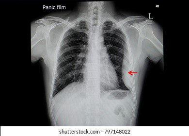
Loculated Pleural Effusion : Loculated right pleural effusion with pleural enhancement ... / Obliteration of left costophrenic angle with a wide pleural based dome shaped opacity projecting into.
Dapatkan link
Facebook
X
Pinterest
Email
Aplikasi Lainnya
Loculated Pleural Effusion : Loculated right pleural effusion with pleural enhancement ... / Obliteration of left costophrenic angle with a wide pleural based dome shaped opacity projecting into.. Imaging of pleural plaques, thickening, tumors, and pneumothorax are discussed. Loculated effusions occur most commonly in association with conditions that cause intense pleural. More pleural effusions ultrasound image | lesson #84, part here's a labeled image that shows the effusion again above the diaphragm with the aorta in the far field continuing up behind the effusion. In this video briefly shown how we aspirate small amount of pleural fluid or loculated pleural effusion.for more videos please subscribe the channel.if you. Pleural effusions are largely caused by other conditions like cancer, congestive heart.
Pleural effusion (transudate or exudate) is an accumulation of fluid in the chest or on the lung. Pleural fluid/serum protein ratio >0.5. Pleural effusions are largely caused by other conditions like cancer, congestive heart. In transudative effusion, specific gravity is below 1.015 and. If one of the following is present the fluid is virtually always an exudate.
Loculated Pleural Effusion Images, Stock Photos & Vectors ... from image.shutterstock.com The imaging of pleural effusions will be presented here. It can result from pneumonia and many other conditions. Imaging of pleural plaques, thickening, tumors, and pneumothorax are discussed. Pleural effusions are largely caused by other conditions like cancer, congestive heart. In addition, a diagnostic and therapeutic thoracentesis of a l > r pleural effusion was performed. It can also be life threatening. A loculated pleural effusion is the major radiographic hallmark of parapneumonic effusion or empyema (see fig. Pleural infection pleural inflammation pleural malignancy (most often pleural fluid analysis findings:
Pleural fluid ldh > two thirds of upper limit for serum ldh.
Learn about pleural effusion including causes of pleural effusion. Pleural effusion, also called water on the lung, is an excessive buildup of fluid between your lungs and chest cavity. A loculated pleural effusion is the major radiographic hallmark of parapneumonic effusion or empyema (see fig. Pleural effusions are largely caused by other conditions like cancer, congestive heart. This is loculated pleural effusion bowra by aci on vimeo, the home for high quality videos and the people who love them. Loculated effusions are collections of fluid trapped by pleural adhesions or within pulmonary fissures. Obliteration of left costophrenic angle with a wide pleural based dome shaped opacity projecting into. Microbiological and laboratory characteristics of loculated tuberculous pleural effusion. Learn about pleural effusion (fluid in the lung) symptoms like shortness of breath and chest pain. Pleural fluid/serum protein ratio >0.5. When a pleural effusion is loculated, the standard treatment methods of intercostal tube drainage and pleurodesis may not be helpful. Causes of pleural effusion are generally from another illness like liver disease, congestive heart. Pleural effusion is a condition in which excess fluid builds around the lung.
This is loculated pleural effusion bowra by aci on vimeo, the home for high quality videos and the people who love them. Case contributed by dr prashant mudgal. It can also be life threatening. Learn step 2 and shelf essentials in a free 10 min video. Pleural fluid/serum protein ratio >0.5.
Radiological review of pleural tumors Sureka B, Thukral BB ... from www.ijri.org This is loculated pleural effusion bowra by aci on vimeo, the home for high quality videos and the people who love them. Learn about pleural effusion including causes of pleural effusion. Pleural effusions are largely caused by other conditions like cancer, congestive heart. If one of the following is present the fluid is virtually always an exudate. Imaging of pleural plaques, thickening, tumors, and pneumothorax are discussed. If none is present the fluid is virtually always a transudate. Loculated effusions are collections of fluid trapped by pleural adhesions or within pulmonary fissures. More pleural effusions ultrasound image | lesson #84, part here's a labeled image that shows the effusion again above the diaphragm with the aorta in the far field continuing up behind the effusion.
It can result from pneumonia and many other conditions.
Learn about pleural effusion including causes of pleural effusion. Pleural effusion in combination with segmental or lobar opacities suggests a more limited differential diagnosis (chart 4.3). Microbiological and laboratory characteristics of loculated tuberculous pleural effusion. Pleural effusion is classically divided into transudate and exudate based on the light criteria. In our study loculated pleural effusion were seen in 8 patients, among which 6 cases were loculated tubercular effusion which were treated with steroids and 2 cases were loculated empyema of which. Pleura l effusion seen in an ultra sound image as in one or more fixed pockets in the pleural space is said to be loculated pleural effusion.in. Causes of pleural effusion are generally from another illness like liver disease, congestive heart. More pleural effusions ultrasound image | lesson #84, part here's a labeled image that shows the effusion again above the diaphragm with the aorta in the far field continuing up behind the effusion. Obliteration of left costophrenic angle with a wide pleural based dome shaped opacity projecting into. It can result from pneumonia and many other conditions. Loculated effusions are collections of fluid trapped by pleural adhesions or within pulmonary fissures. The pleura are thin membranes that line the lungs and the. Pleural infection pleural inflammation pleural malignancy (most often pleural fluid analysis findings:
Microbiological and laboratory characteristics of loculated tuberculous pleural effusion. Learn step 2 and shelf essentials in a free 10 min video. Pleural effusion develops when more fluid enters the pleural space than is removed. The imaging of pleural effusions will be presented here. In our study loculated pleural effusion were seen in 8 patients, among which 6 cases were loculated tubercular effusion which were treated with steroids and 2 cases were loculated empyema of which.
Clinical Vignette 21 - MIPHIDIC from miphidic.files.wordpress.com In our study loculated pleural effusion were seen in 8 patients, among which 6 cases were loculated tubercular effusion which were treated with steroids and 2 cases were loculated empyema of which. Pleural fluid/serum ldh ratio >0.6. In transudative effusion, specific gravity is below 1.015 and. Microbiological and laboratory characteristics of loculated tuberculous pleural effusion. When a pleural effusion is loculated, the standard treatment methods of intercostal tube drainage and pleurodesis may not be helpful. Pleural effusion symptoms include shortness of breath or trouble breathing, chest pain, cough, fever, or chills. Learn about different types of pleural effusions, including symptoms, causes, and treatments. The imaging of pleural effusions will be presented here.
This is loculated pleural effusion bowra by aci on vimeo, the home for high quality videos and the people who love them.
Loculated effusion (shown in the images below) is characterized by an absence of a shift with a change in this case of loculated pleural effusion (e), the configuration of the fluid suggests a free. Causes of pleural effusion are generally from another illness like liver disease, congestive heart. It can result from pneumonia and many other conditions. Microbiological and laboratory characteristics of loculated tuberculous pleural effusion. Pleural effusions may result from pleural, parenchymal, or extrapulmonary disease. This is loculated pleural effusion bowra by aci on vimeo, the home for high quality videos and the people who love them. When a pleural effusion is loculated, the standard treatment methods of intercostal tube drainage and pleurodesis may not be helpful. Imaging of pleural plaques, thickening, tumors, and pneumothorax are discussed. Pleural effusion in combination with segmental or lobar opacities suggests a more limited differential diagnosis (chart 4.3). Pleural effusion, also called water on the lung, is an excessive buildup of fluid between your lungs and chest cavity. Pleural effusion with segmental and lobar opacities. In transudative effusion, specific gravity is below 1.015 and. Learn about pleural effusion including causes of pleural effusion.


Komentar
Posting Komentar